Bradford Protein Assay
Goal: To estimate total protein content in a small
sample of a complex substance
examples:
- milk
- homogenized foods (meat, lettuce, beans, etc)
History
Biuret, Lowry (1956) and Bradford (1976) are the most popular tests (assays)
for estimating total protein content of a mixture.
In this lab-lesson, we use the Bio-Rad Bradford Protein Assay reagent.
Coomassie Blue, under acidic conditions, forms a microprecipitate with many
(but not all) proteins. In the process, a spectral shift of light absorbance
moves from a maximum in the red region to the blue region, i.e., the pigment
turns blue in the presence of protein.
Pierce and Sigma also market reagent for the Bradford protein assay; however,
the Bio-Rad reagent generates colors of blue that are easier to discern with
our eyes alone.
Dr. Toby has been using the Bradford Protein Assay for her research since it
was published in Analytical Biochemistry in 1976 and has been teaching high
school students the protein assay using the Bio-Rad reagent since 1985.
Manual for Teaching (Adobe acrobat
format .pdf, 104K, 32 pages)
Manufacturers Manual
(Bio-Rad link also in .pdf format)
Images for troubleshooting
|
|
Volumes in the 24-well dish. The middle is the correct volume
for the protein assay. |
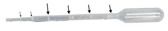 |
Transfer pipette.
Large arrows point to increments of 0.25 ml.
|
|
1 ml pipette. Use this with the blue pi-pump for conducting
the protein assay.
Do not push the top (on the left in this image) all the way into the pi-pump.
Some of that top section should still be visible. Hold the pipette between
the double line of safety and the top (aseptic handling). Note that the
major numbered divisions are 0.1 ml. Those are the divisions to use for
conducting the assay---not the tinier divisions.
|
|
|
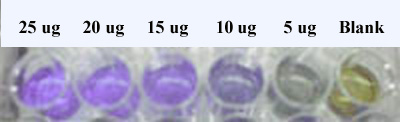
Colors of a typical Standard Curve.
The Blank (right) has no protein added. Only one row of duplicates is
shown.
|
.jpg)