Individual Cell Images --- What fields of cells look like in the microscope --- Parts of the Cell
Click on the images for links to more ...

How Microscopes Work

Which Is Bigger?

Onion Root Tips: No microscopes needed to view and collect data from these excellent slide images (Kansas University Medical Center)

cheek cell
http://www.durr.demon.co.uk/ algae%20&%20protozoa%202.html

sickled cell --- red blood cells --- normal cell

source http://www.sicklecellfoundationofalberta.org/
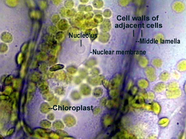
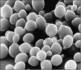
Elodea cells
http://www.biosci.ohio-state.edu/~plantbio/images/images%20from%20Lima/chlorop.jpg

Bacteria cells
http://www.life.umd.edu/classroom/bsci124/img/bacteria.jpg

Yeast cells
http://csm.jmu.edu/biology/danie2jc/bio220/homepage.htm


Paramecium
http://www.microscopy-uk.org.uk/mag/artmar99/marimg99.html

http://www.sp.uconn.edu/~bi107vc/images/cell/sperm+egg.JPG

Amoeba
http://www.microscopy-uk.org.uk/mag/artsep01/amoeba.html

http://vis-pc.plantbio.ohiou.edu/algaeimage/pages/Acetabularia.html

http://www.wellesley.edu/Biology/Courses/108/108.html

Zebrafish egg and embryo BAR is 250 µm
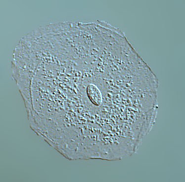
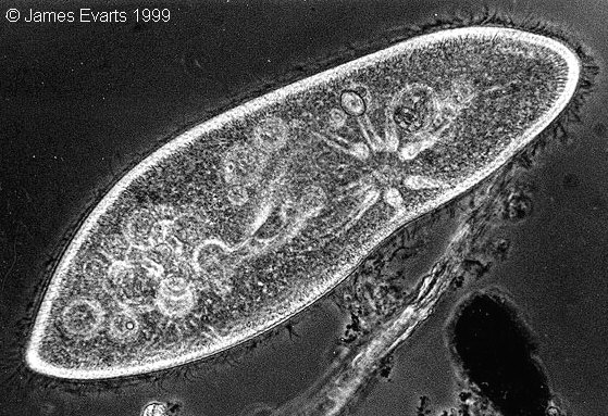
http://zfin.org/zf_info/zfbook/stages/figs/fig3.html

comparative fields --- http://www.usoe.k12.ut.us/curr/science/sciber00/7th/cells/sciber/cellphot.htm

http://mimp.mems.cmu.edu/~ordofmag/which%20is%20bigger%202.htm

Virtual Cell

http://www.veso.no/dnavaccine/partner1.htm


Stem Cells in Culture


Karyotype

Want even more?

Tutorial/quiz on histology